Diag Image: A Comprehensive Guide to Modern Diagnostic Radiology
The world of healthcare is rapidly evolving, and at the forefront of this transformation is diagnostic radiology. This field plays a crucial role in identifying medical conditions through advanced imaging techniques. But what exactly does it entail? Enter Diag Image, a term synonymous with cutting-edge technology that helps healthcare professionals visualize what’s happening inside the human body without invasive procedures.
With each passing year, innovations reshape how we understand diseases. From X-rays to MRIs and CT scans, these tools have become vital in diagnosing illnesses early and accurately. As patients seek answers faster than ever before, understanding diagnostic imaging has never been more critical.
In this blog post, we’ll explore everything you need to know about Diag Image—its evolution, various types of tools used today, their benefits and limitations across different medical fields—and how recent advancements are paving the way for future developments in healthcare. Join us as we dive into the fascinating realm of modern diagnostic radiology!
What is Diag Image?
Diag Image refers to the integration of advanced imaging technology in diagnostic radiology. It encompasses various techniques that allow healthcare professionals to visualize the internal structures of the body.
This innovative approach is crucial for accurate diagnosis and treatment planning. By utilizing sophisticated equipment, Diag Image enhances clinicians’ abilities to detect diseases at an early stage.
Techniques under this umbrella include X-rays, MRI scans, CT scans, and ultrasound imaging. Each serves a unique purpose and contributes distinct information about patients’ health conditions.
Moreover, Diag Image not only improves patient outcomes but also streamlines workflows within medical facilities. As technology evolves, so does its potential impact on diagnostics in modern medicine.
Evolution of Diagnostic Imaging Techniques
The journey of diagnostic imaging has been remarkable. It began in the late 19th century with Wilhelm Conrad Röntgen’s discovery of X-rays. This groundbreaking innovation allowed for non-invasive visualization of bones, changing medicine forever.
As technology advanced, so did imaging techniques. The introduction of ultrasound in the mid-20th century offered a new way to visualize soft tissues and monitor fetal development without radiation exposure.
Later, computed tomography (CT) revolutionized diagnostics by combining X-ray images from multiple angles to create detailed cross-sectional views of the body. Magnetic resonance imaging (MRI) followed closely behind, using powerful magnets and radio waves for even finer details.
Digital advancements opened doors to more sophisticated tools like positron emission tomography (PET), merging functional and anatomical information into one image. Each evolution enhanced precision and safety while paving the way for innovative approaches in modern healthcare delivery.
Types of Diagnostic Imaging Tools in Modern Medicine
Diagnostic imaging tools are essential in modern medicine, providing clarity and insights into the human body. Each technique has its unique strengths.
X-rays remain one of the most common methods. They offer quick visualization of bones and certain tissues, making them invaluable for identifying fractures or infections.
Computed Tomography (CT) scans provide cross-sectional images, revealing detailed views of internal structures. This tool is particularly effective in diagnosing conditions like tumors or organ abnormalities.
Magnetic Resonance Imaging (MRI) takes advantage of strong magnets and radio waves to produce high-resolution images. It excels at visualizing soft tissues, making it ideal for brain disorders and joint injuries.
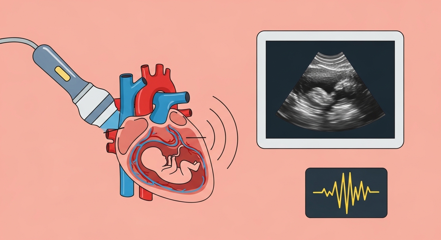
Ultrasound uses sound waves to create real-time images. It’s often employed during pregnancy but also plays a role in assessing organs such as the heart and liver.
Each imaging tool contributes uniquely to patient assessment, enhancing diagnostic accuracy across various medical fields.
Benefits and Limitations of Each Imaging Technique
Every imaging technique has its own set of advantages and drawbacks. X-rays are quick and widely available, making them ideal for initial assessments. However, they expose patients to radiation, which raises safety concerns.
MRI scans offer detailed images without radiation exposure. They excel in soft tissue evaluation but can be time-consuming and uncomfortable due to the enclosed space.
CT scans provide rapid results with high-resolution images. They’re excellent for trauma cases but also involve higher radiation doses compared to X-rays.
Ultrasound is safe, cost-effective, and real-time visualized; it’s particularly beneficial in obstetrics. The downside? Its effectiveness can be limited by patient body type or operator skill.
Nuclear medicine techniques like PET scans deliver unique insights into metabolic functions but may require longer preparation times and introduce radioactive tracers into the body. Each method brings something valuable while presenting certain limitations that must be considered carefully in clinical practice.
Common Uses of Diagnostic Imaging in Various Medical Fields
Diagnostic imaging plays a crucial role across various medical fields, providing essential insights into patient conditions.
In oncology, it aids in detecting tumors early and monitoring treatment response. Techniques like MRI and PET scans are invaluable for identifying malignancies with precision.
Cardiology relies heavily on echocardiograms and angiography to visualize heart structures and blood flow. These images help diagnose conditions such as blockages or congenital heart defects.
Orthopedics benefits from X-rays and CT scans that reveal fractures or joint issues. Surgeons utilize these images for accurate pre-operative planning.
Neurology employs advanced imaging like MRIs to assess brain injuries, strokes, or neurological disorders. This information is critical for developing effective treatment plans.
Additionally, pediatrics uses ultrasound extensively for fetal assessments during pregnancy, ensuring the health of both mother and child remains a priority even before birth. Each specialty harnesses diagnostic imaging’s power to enhance patient care dramatically.
Latest Technological Advancements in Diagnostic Radiology
Recent advancements in diagnostic radiology are transforming patient care. Artificial intelligence (AI) is leading the charge, enhancing image analysis and interpretation. Algorithms can now identify abnormalities faster than ever before, allowing for quicker diagnoses.
3D imaging technology has also made significant strides. This innovation provides a more detailed view of internal structures, aiding surgeons in planning complex procedures with precision.
Moreover, portable imaging devices are becoming increasingly popular. These tools offer flexibility and convenience, enabling healthcare providers to conduct scans at patients’ bedsides or remote locations without compromising quality.
Tele-radiology is another exciting development. It allows radiologists to share images and reports over long distances efficiently. This capability improves access to expert opinions and speeds up treatment decisions for patients everywhere.
The integration of these technologies paves the way for more personalized medicine by tailoring treatments based on accurate diagnostics.
Future Scope and Potential Impact of Diagnostic Imaging on Healthcare
The future of diagnostic imaging promises remarkable advancements that could transform healthcare. As artificial intelligence continues to evolve, we can expect smarter algorithms capable of analyzing images with unprecedented accuracy. This will enhance the detection of diseases at earlier stages.
Moreover, integration with telemedicine is on the rise. Patients from remote areas will receive timely diagnoses without needing to travel long distances for specialized care. This accessibility could bridge gaps in healthcare access.
3D printing technology is also making waves. Custom implants and prosthetics derived from imaging data may soon become commonplace, leading to more personalized treatment plans.
Additionally, molecular imaging techniques are set to advance precision medicine further. Tailoring treatments based on individual responses becomes feasible as we gain deeper insights into disease mechanisms through these innovative methods.
With ongoing research and development, diagnostic imaging stands ready to play a pivotal role in shaping a more effective and responsive healthcare system.
Conclusion
Diagnostic radiology has come a long way since its inception. With techniques evolving rapidly, the field of Diag Image stands at the forefront of medical technology. The diverse range of imaging tools available today—each with unique benefits and limitations—has transformed how healthcare professionals diagnose and treat patients.
From X-rays to MRI scans, these diagnostic methods have opened new avenues for understanding complex health conditions. Their applications stretch across various medical fields, aiding in everything from emergency medicine to oncology.
As technological advancements continue to unfold, we can expect even more precision and efficiency in diagnostic imaging. Innovations such as artificial intelligence are poised to enhance image analysis further, making diagnoses faster and more accurate.